Figure 2
Painful unilateral gynecomastia with identification of the cause of the pain: A case report
Charles M Lombard* and Peter L Naruns
Published: 31 December, 2021 | Volume 5 - Issue 1 | Pages: 034-036
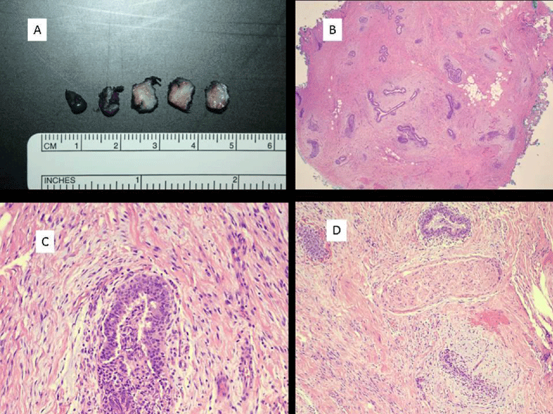
Figure 2:
Gross appearance of gynecomastia with a circumscribed nodule of edematous fibrous tissue. B) Low power image of gynecomastia with the proliferation of glandular and stromal elements (hematoxylin and eosin; 20 x magnification). C) Image of gynecomastia showing the epithelial proliferation without atypia and the surrounding stromal proliferation with associated stromal edema (hematoxylin and eosin, 200x magnification). D) Image showing peripheral nerve intimately admixed with gynecomastia-associated proliferation and compressed between proliferation with associated edema (hematoxylin and eosin, 100 x magnification).
Read Full Article HTML DOI: 10.29328/journal.apcr.1001026 Cite this Article Read Full Article PDF
More Images
Similar Articles
-
MicroRNA Therapeutics in Triple Negative Breast CancerSarmistha Mitra*. MicroRNA Therapeutics in Triple Negative Breast Cancer . . 2017 doi: 10.29328/journal.hjpcr.1001003; 1: 009-017
-
A great mimicker of Bone Secondaries: Brown Tumors, presenting with a Degenerative Lumber Disc like painZuhal Bayramoglu*,Ravza Yılmaz,Aysel Bayram. A great mimicker of Bone Secondaries: Brown Tumors, presenting with a Degenerative Lumber Disc like pain. . 2017 doi: 10.29328/journal.hjpcr.1001004; 1: 018-023
-
Amyotropyc Lateral Sclerosis and Endogenous -Esogenous Toxicological Movens: New model to verify other Pharmacological StrategiesMauro Luisetto*,Behzad Nili-Ahmadabadi,Nilesh M Meghani,Ghulam Rasool Mashori,Ram Kumar Sahu,Kausar Rehman Khan, Ahmed Yesvi Rafa,Luca Cabianca,Gamal Abdul Hamid, Farhan Ahmad Khan. Amyotropyc Lateral Sclerosis and Endogenous -Esogenous Toxicological Movens: New model to verify other Pharmacological Strategies. . 2018 doi: 10.29328/journal.apcr.1001009; 2: 029-048
-
Receptor pharmacology and other relevant factors in lower urinary tract pathology under a functional and toxicological approach: Instrument to better manage antimicrobials therapyMauro Luisetto*,Naseer Almukhtar,Behzad Nili-Ahmadabadi,Ghulam Rasool Mashori,Kausar Rehman Khan,Ram Kumar Sahu,Farhan Ahmad Khan,Gamal Abdul Hamid,Luca Cabianca. Receptor pharmacology and other relevant factors in lower urinary tract pathology under a functional and toxicological approach: Instrument to better manage antimicrobials therapy . . 2018 doi: 10.29328/journal.apcr.1001010; 2: 049-093
-
The pathogenesis of psoriasis: insight into a complex “Mobius Loop” regulation processYuankuan Jiang,Haiyang Chen,Jiayue Liu,Tianfu Wei,Peng Ge,Jialin Qu*,Jingrong Lin. The pathogenesis of psoriasis: insight into a complex “Mobius Loop” regulation process. . 2021 doi: 10.29328/journal.apcr.1001024; 5: 020-025
-
Painful unilateral gynecomastia with identification of the cause of the pain: A case reportCharles M Lombard*,Peter L Naruns. Painful unilateral gynecomastia with identification of the cause of the pain: A case report. . 2021 doi: 10.29328/journal.apcr.1001026; 5: 034-036
-
Immune-mediated neuropathy related to bortezomib in a patient with multiple myelomaSusanne Koeppen*,Jörg Hense,Kay Wilhelm Nolte,Joachim Weis. Immune-mediated neuropathy related to bortezomib in a patient with multiple myeloma. . 2022 doi: 10.29328/journal.apcr.1001028; 6: 001-004
-
Post-operative agranulocytosis caused by intravenous cefazolin: A case report with a discussion of the pathogenesisCharles M Lombard*,Jiali Li,Bijayee Shrestha. Post-operative agranulocytosis caused by intravenous cefazolin: A case report with a discussion of the pathogenesis. . 2022 doi: 10.29328/journal.apcr.1001030; 6: 009-012
-
Harmonizing Artificial Intelligence Governance; A Model for Regulating a High-risk Categories and Applications in Clinical Pathology: The Evidence and some ConcernsMaxwell Omabe*. Harmonizing Artificial Intelligence Governance; A Model for Regulating a High-risk Categories and Applications in Clinical Pathology: The Evidence and some Concerns. . 2024 doi: 10.29328/journal.apcr.1001040; 8: 001-005
-
The Accuracy of pHH3 in Meningioma Grading: A Single Institution StudyMansouri Nada1, Yaiche Rahma*, Takout Khouloud, Gargouri Faten, Tlili Karima, Rachdi Mohamed Amine, Ammar Hichem, Yedeas Dahmani, Radhouane Khaled, Chkili Ridha, Msakni Issam, Laabidi Besma. The Accuracy of pHH3 in Meningioma Grading: A Single Institution Study. . 2024 doi: 10.29328/journal.apcr.1001041; 8: 006-011
Recently Viewed
-
Methodology for Studying Combustion of Solid Rocket Propellants using Artificial Neural NetworksVictor Abrukov*, Weiqiang Pang, Darya Anufrieva. Methodology for Studying Combustion of Solid Rocket Propellants using Artificial Neural Networks. Ann Adv Chem. 2024: doi: 10.29328/journal.aac.1001048; 8: 001-007
-
A Study on Potential Feed Sources to Boost Guppy Fish, Poecilia reticulata ProductivityCV Vedhavarshini, Swetha A, Harini Sri M, Kaviya K, Ann Suji H, Deivesigamani B*. A Study on Potential Feed Sources to Boost Guppy Fish, Poecilia reticulata Productivity. Ann Adv Chem. 2024: doi: 10.29328/journal.aac.1001049; 8: 008-011
-
Using Isomets as a Foundation, a Connection Factor between Nucleation and Atomic PhysicsEsraa Fareed Saeed*. Using Isomets as a Foundation, a Connection Factor between Nucleation and Atomic Physics. Ann Adv Chem. 2024: doi: 10.29328/journal.aac.1001050; 8: 012-018
-
Techno-econophysics’ Fractal Involving of Exergy RemarksWidastra Hidajatullah Maksoed*. Techno-econophysics’ Fractal Involving of Exergy Remarks. Ann Adv Chem. 2024: doi: 10.29328/journal.aac.1001051; 8: 017-020
-
Comparative Studies of Diclofenac Sodium (NSAID) Adsorption on Wheat (Triticum aestivum) Bran and Groundnut (Arachis hypogaea) Shell Powder using Vertical and Sequential Bed ColumnNeha Dhiman*. Comparative Studies of Diclofenac Sodium (NSAID) Adsorption on Wheat (Triticum aestivum) Bran and Groundnut (Arachis hypogaea) Shell Powder using Vertical and Sequential Bed Column. Ann Adv Chem. 2024: doi: 10.29328/journal.aac.1001052; 8: 021-029
Most Viewed
-
Evaluation of Biostimulants Based on Recovered Protein Hydrolysates from Animal By-products as Plant Growth EnhancersH Pérez-Aguilar*, M Lacruz-Asaro, F Arán-Ais. Evaluation of Biostimulants Based on Recovered Protein Hydrolysates from Animal By-products as Plant Growth Enhancers. J Plant Sci Phytopathol. 2023 doi: 10.29328/journal.jpsp.1001104; 7: 042-047
-
Sinonasal Myxoma Extending into the Orbit in a 4-Year Old: A Case PresentationJulian A Purrinos*, Ramzi Younis. Sinonasal Myxoma Extending into the Orbit in a 4-Year Old: A Case Presentation. Arch Case Rep. 2024 doi: 10.29328/journal.acr.1001099; 8: 075-077
-
Feasibility study of magnetic sensing for detecting single-neuron action potentialsDenis Tonini,Kai Wu,Renata Saha,Jian-Ping Wang*. Feasibility study of magnetic sensing for detecting single-neuron action potentials. Ann Biomed Sci Eng. 2022 doi: 10.29328/journal.abse.1001018; 6: 019-029
-
Pediatric Dysgerminoma: Unveiling a Rare Ovarian TumorFaten Limaiem*, Khalil Saffar, Ahmed Halouani. Pediatric Dysgerminoma: Unveiling a Rare Ovarian Tumor. Arch Case Rep. 2024 doi: 10.29328/journal.acr.1001087; 8: 010-013
-
Physical activity can change the physiological and psychological circumstances during COVID-19 pandemic: A narrative reviewKhashayar Maroufi*. Physical activity can change the physiological and psychological circumstances during COVID-19 pandemic: A narrative review. J Sports Med Ther. 2021 doi: 10.29328/journal.jsmt.1001051; 6: 001-007

HSPI: We're glad you're here. Please click "create a new Query" if you are a new visitor to our website and need further information from us.
If you are already a member of our network and need to keep track of any developments regarding a question you have already submitted, click "take me to my Query."


