Figure 1
Uterine precursor lesions in patients with incidental nodal lymphangioleiomyomatosis: A report of 4 cases
Charles M Lombard*
Published: 14 December, 2020 | Volume 4 - Issue 1 | Pages: 001-004.
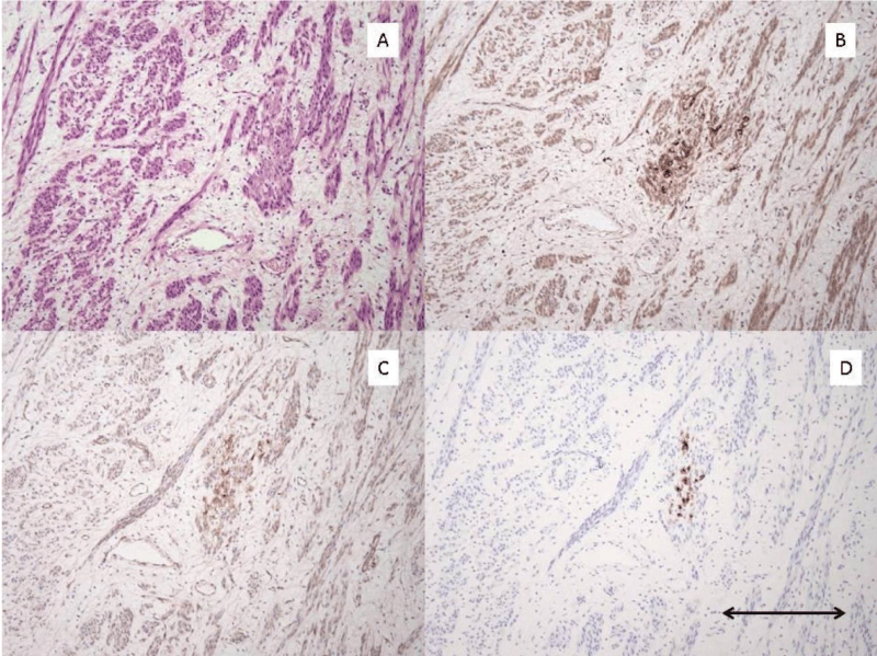
Figure 1:
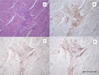
Pre-LAM uterine lesion pattern #1. A): H/E stain of area with Pre-LAM lesion composed of cluster of small epithelioid appearing cells. Lesional cells are inapparent on this stain. B): Cathepsin K IHC stain shows strong positive staining of the small cluster of epithelioid cells. C): Beta-catenin IHC stain shows moderate staining of the small cluster of epithelioid cells. D): HMB-45 IHC stain shows strong positive staining of the small cluster of epithelioid cells. All photomicrographs taken at 100x magnification. Scale bar in lower right corner measures 0.2 mm.
Read Full Article HTML DOI: 10.29328/journal.apcr.1001016 Cite this Article Read Full Article PDF
More Images
Similar Articles
-
MicroRNA Therapeutics in Triple Negative Breast CancerSarmistha Mitra*. MicroRNA Therapeutics in Triple Negative Breast Cancer . . 2017 doi: 10.29328/journal.hjpcr.1001003; 1: 009-017
-
Amyotropyc Lateral Sclerosis and Endogenous -Esogenous Toxicological Movens: New model to verify other Pharmacological StrategiesMauro Luisetto*,Behzad Nili-Ahmadabadi,Nilesh M Meghani,Ghulam Rasool Mashori,Ram Kumar Sahu,Kausar Rehman Khan, Ahmed Yesvi Rafa,Luca Cabianca,Gamal Abdul Hamid, Farhan Ahmad Khan. Amyotropyc Lateral Sclerosis and Endogenous -Esogenous Toxicological Movens: New model to verify other Pharmacological Strategies. . 2018 doi: 10.29328/journal.apcr.1001009; 2: 029-048
-
Receptor pharmacology and other relevant factors in lower urinary tract pathology under a functional and toxicological approach: Instrument to better manage antimicrobials therapyMauro Luisetto*,Naseer Almukhtar,Behzad Nili-Ahmadabadi,Ghulam Rasool Mashori,Kausar Rehman Khan,Ram Kumar Sahu,Farhan Ahmad Khan,Gamal Abdul Hamid,Luca Cabianca. Receptor pharmacology and other relevant factors in lower urinary tract pathology under a functional and toxicological approach: Instrument to better manage antimicrobials therapy . . 2018 doi: 10.29328/journal.apcr.1001010; 2: 049-093
-
Uterine precursor lesions in patients with incidental nodal lymphangioleiomyomatosis: A report of 4 casesCharles M Lombard*. Uterine precursor lesions in patients with incidental nodal lymphangioleiomyomatosis: A report of 4 cases . . 2020 doi: 10.29328/journal.apcr.1001016; 4: 001-004.
-
The pathogenesis of psoriasis: insight into a complex “Mobius Loop” regulation processYuankuan Jiang,Haiyang Chen,Jiayue Liu,Tianfu Wei,Peng Ge,Jialin Qu*,Jingrong Lin. The pathogenesis of psoriasis: insight into a complex “Mobius Loop” regulation process. . 2021 doi: 10.29328/journal.apcr.1001024; 5: 020-025
-
Immune-mediated neuropathy related to bortezomib in a patient with multiple myelomaSusanne Koeppen*,Jörg Hense,Kay Wilhelm Nolte,Joachim Weis. Immune-mediated neuropathy related to bortezomib in a patient with multiple myeloma. . 2022 doi: 10.29328/journal.apcr.1001028; 6: 001-004
-
Post-operative agranulocytosis caused by intravenous cefazolin: A case report with a discussion of the pathogenesisCharles M Lombard*,Jiali Li,Bijayee Shrestha. Post-operative agranulocytosis caused by intravenous cefazolin: A case report with a discussion of the pathogenesis. . 2022 doi: 10.29328/journal.apcr.1001030; 6: 009-012
-
Harmonizing Artificial Intelligence Governance; A Model for Regulating a High-risk Categories and Applications in Clinical Pathology: The Evidence and some ConcernsMaxwell Omabe*. Harmonizing Artificial Intelligence Governance; A Model for Regulating a High-risk Categories and Applications in Clinical Pathology: The Evidence and some Concerns. . 2024 doi: 10.29328/journal.apcr.1001040; 8: 001-005
-
The Accuracy of pHH3 in Meningioma Grading: A Single Institution StudyMansouri Nada1, Yaiche Rahma*, Takout Khouloud, Gargouri Faten, Tlili Karima, Rachdi Mohamed Amine, Ammar Hichem, Yedeas Dahmani, Radhouane Khaled, Chkili Ridha, Msakni Issam, Laabidi Besma. The Accuracy of pHH3 in Meningioma Grading: A Single Institution Study. . 2024 doi: 10.29328/journal.apcr.1001041; 8: 006-011
Recently Viewed
-
Cystoid Macular Oedema Secondary to Bimatoprost in a Patient with Primary Open Angle GlaucomaKonstantinos Kyratzoglou*,Katie Morton. Cystoid Macular Oedema Secondary to Bimatoprost in a Patient with Primary Open Angle Glaucoma. Int J Clin Exp Ophthalmol. 2025: doi: 10.29328/journal.ijceo.1001059; 9: 001-003
-
Metastatic Brain Melanoma: A Rare Case with Review of LiteratureNeha Singh,Gaurav Raj,Akshay Kumar,Deepak Kumar Singh,Shivansh Dixit,Kaustubh Gupta*. Metastatic Brain Melanoma: A Rare Case with Review of Literature. J Radiol Oncol. 2025: doi: ; 9: 050-053
-
Depression as a civilization-deformed adaptation and defence mechanismBohdan Wasilewski*,Olha Yourtsenyuk,Eugene Egan. Depression as a civilization-deformed adaptation and defence mechanism. Insights Depress Anxiety. 2020: doi: 10.29328/journal.ida.1001013; 4: 008-011
-
Drinking-water Quality Assessment in Selective Schools from the Mount LebanonWalaa Diab, Mona Farhat, Marwa Rammal, Chaden Moussa Haidar*, Ali Yaacoub, Alaa Hamzeh. Drinking-water Quality Assessment in Selective Schools from the Mount Lebanon. Ann Civil Environ Eng. 2024: doi: 10.29328/journal.acee.1001061; 8: 018-024
-
Rapid Microbial Growth in Reusable Drinking Water BottlesQishan Liu*,Hongjun Liu. Rapid Microbial Growth in Reusable Drinking Water Bottles. Ann Civil Environ Eng. 2017: doi: 10.29328/journal.acee.1001007; 1: 055-062
Most Viewed
-
Evaluation of Biostimulants Based on Recovered Protein Hydrolysates from Animal By-products as Plant Growth EnhancersH Pérez-Aguilar*, M Lacruz-Asaro, F Arán-Ais. Evaluation of Biostimulants Based on Recovered Protein Hydrolysates from Animal By-products as Plant Growth Enhancers. J Plant Sci Phytopathol. 2023 doi: 10.29328/journal.jpsp.1001104; 7: 042-047
-
Sinonasal Myxoma Extending into the Orbit in a 4-Year Old: A Case PresentationJulian A Purrinos*, Ramzi Younis. Sinonasal Myxoma Extending into the Orbit in a 4-Year Old: A Case Presentation. Arch Case Rep. 2024 doi: 10.29328/journal.acr.1001099; 8: 075-077
-
Feasibility study of magnetic sensing for detecting single-neuron action potentialsDenis Tonini,Kai Wu,Renata Saha,Jian-Ping Wang*. Feasibility study of magnetic sensing for detecting single-neuron action potentials. Ann Biomed Sci Eng. 2022 doi: 10.29328/journal.abse.1001018; 6: 019-029
-
Pediatric Dysgerminoma: Unveiling a Rare Ovarian TumorFaten Limaiem*, Khalil Saffar, Ahmed Halouani. Pediatric Dysgerminoma: Unveiling a Rare Ovarian Tumor. Arch Case Rep. 2024 doi: 10.29328/journal.acr.1001087; 8: 010-013
-
Physical activity can change the physiological and psychological circumstances during COVID-19 pandemic: A narrative reviewKhashayar Maroufi*. Physical activity can change the physiological and psychological circumstances during COVID-19 pandemic: A narrative review. J Sports Med Ther. 2021 doi: 10.29328/journal.jsmt.1001051; 6: 001-007

HSPI: We're glad you're here. Please click "create a new Query" if you are a new visitor to our website and need further information from us.
If you are already a member of our network and need to keep track of any developments regarding a question you have already submitted, click "take me to my Query."


