Abstract
Case Report
The identification of the true nature of pseudofungus structures as polyurethane catheter fragments
Charles M Lombard*
Published: 04 January, 2022 | Volume 6 - Issue 1 | Pages: 005-008
Pseudofungus structures in lymph node tissues have been reported on multiple occasions. Despite a variety of investigative tests including histochemical special stains and energy dispersive spectral analysis, the underlying nature and origin of these pseudofungus structures has never been clearly defined. The most common hypothesis suggests that they represent collagen fibers that become coated with iron and calcium. Herein, evidence is given that the pseudofungus structures identified in the lymph node tissues represent fragments of polyurethane catheters. The evidence includes both a comparison of these pseudofungus structures to fragments of polyurethane well documented in the literature and a comparison of polyurethane catheter scrapings to the pseudofungus structures identified in the literature. In both of these comparisons, the morphology of the polyurethane fragments are identical to the pseudofungus structures. This is the first definitive report identifying polyurethane catheter fragments as representing the true nature and etiology of pseudofungus structures in lymph node tissues.
Read Full Article HTML DOI: 10.29328/journal.apcr.1001029 Cite this Article Read Full Article PDF
Keywords:
Pseudofungus; Pseudofungi; Polyurethane catheter fragments; Pathogenesis; Pathology
References
- Connelly J, Cartwright J. Pseudofungi in a lymph node: A case report with energy dispersive x-ray elemental analysis. Arch Pathol Lab Med. 1991; 115: 1166-1168. PubMed: https://pubmed.ncbi.nlm.nih.gov/1747037/
- Teague MD, Tham KT. Hyphalike pseudofungus in the lymph node. Arch Pathol Lab Med. 1994; 118: 95-96. PubMed: https://pubmed.ncbi.nlm.nih.gov/8285843/ b
- Kim H, Koo JS, Shim H, et al. Pseudofungi associated with a granulomatous response in a lymph node: A case report. The Korean J of Pathol. 2004; 38: 64-67.
- Song ES, Kahn AG, Shin KK, Ro JY. Pseudofungi in pericolic lymph nodes. Arch Pathol Lab Med. 2005; 129: e97-e100. PubMed: https://pubmed.ncbi.nlm.nih.gov/15794699/
- Singh S, Manivel JC, Pambuccian SE. Pseudofungi: coral shapes and Bamboo sticks in lymph node sinuses. Int J Surg Pathol. 2010; 18: 68-69. PubMed: https://pubmed.ncbi.nlm.nih.gov/19748899/
- Lyapichev KA, Agarwal AA, Rosenberg AE, Chapman JR. Pseudofungi: A diagnostic pitfall. Int J Surg Pathol. 2016; 24: 528-531. PubMed: https://pubmed.ncbi.nlm.nih.gov/27106780/
- Nigdelioglu R, Biemer J, Pambuccian SE. Liesegang structures and pseudofungi in a cystic renal cell carcinoma. Diagnostic cytopath. 2018; 46: 888-891. PubMed: https://pubmed.ncbi.nlm.nih.gov/30043493/
- Bilani N, Elson L, Carlson D, Elimimian EB, Nahleh Z. Pseudofungi in an immunocompromised patient with breast cancer and COVID-19. Hindawi Case Reports in Medicine. 2020.
- Lau S, Williams D. PAS positive pseudofungi: a diagnostic pitfall. Pathology. 2021; 53: 542-543. PubMed: https://pubmed.ncbi.nlm.nih.gov/33218738/
- Zhou L, Pittmann W. Distributions organophosphate flame retardants (OPFRs) in 3 dust size fractions from homes and building material markets. Environmen Pollut. 2019; 245: 343-352. PubMed: https://pubmed.ncbi.nlm.nih.gov/30448504/
Figures:
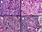
Figure 1
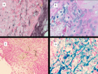
Figure 2
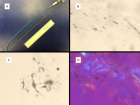
Figure 3
Similar Articles
-
MicroRNA Therapeutics in Triple Negative Breast CancerSarmistha Mitra*. MicroRNA Therapeutics in Triple Negative Breast Cancer . . 2017 doi: 10.29328/journal.hjpcr.1001003; 1: 009-017
-
Amyotropyc Lateral Sclerosis and Endogenous -Esogenous Toxicological Movens: New model to verify other Pharmacological StrategiesMauro Luisetto*,Behzad Nili-Ahmadabadi,Nilesh M Meghani,Ghulam Rasool Mashori,Ram Kumar Sahu,Kausar Rehman Khan, Ahmed Yesvi Rafa,Luca Cabianca,Gamal Abdul Hamid, Farhan Ahmad Khan. Amyotropyc Lateral Sclerosis and Endogenous -Esogenous Toxicological Movens: New model to verify other Pharmacological Strategies. . 2018 doi: 10.29328/journal.apcr.1001009; 2: 029-048
-
Receptor pharmacology and other relevant factors in lower urinary tract pathology under a functional and toxicological approach: Instrument to better manage antimicrobials therapyMauro Luisetto*,Naseer Almukhtar,Behzad Nili-Ahmadabadi,Ghulam Rasool Mashori,Kausar Rehman Khan,Ram Kumar Sahu,Farhan Ahmad Khan,Gamal Abdul Hamid,Luca Cabianca. Receptor pharmacology and other relevant factors in lower urinary tract pathology under a functional and toxicological approach: Instrument to better manage antimicrobials therapy . . 2018 doi: 10.29328/journal.apcr.1001010; 2: 049-093
-
The pathogenesis of psoriasis: insight into a complex “Mobius Loop” regulation processYuankuan Jiang,Haiyang Chen,Jiayue Liu,Tianfu Wei,Peng Ge,Jialin Qu*,Jingrong Lin. The pathogenesis of psoriasis: insight into a complex “Mobius Loop” regulation process. . 2021 doi: 10.29328/journal.apcr.1001024; 5: 020-025
-
The identification of the true nature of pseudofungus structures as polyurethane catheter fragmentsCharles M Lombard*. The identification of the true nature of pseudofungus structures as polyurethane catheter fragments. . 2022 doi: 10.29328/journal.apcr.1001029; 6: 005-008
-
Immune-mediated neuropathy related to bortezomib in a patient with multiple myelomaSusanne Koeppen*,Jörg Hense,Kay Wilhelm Nolte,Joachim Weis. Immune-mediated neuropathy related to bortezomib in a patient with multiple myeloma. . 2022 doi: 10.29328/journal.apcr.1001028; 6: 001-004
-
Post-operative agranulocytosis caused by intravenous cefazolin: A case report with a discussion of the pathogenesisCharles M Lombard*,Jiali Li,Bijayee Shrestha. Post-operative agranulocytosis caused by intravenous cefazolin: A case report with a discussion of the pathogenesis. . 2022 doi: 10.29328/journal.apcr.1001030; 6: 009-012
-
Harmonizing Artificial Intelligence Governance; A Model for Regulating a High-risk Categories and Applications in Clinical Pathology: The Evidence and some ConcernsMaxwell Omabe*. Harmonizing Artificial Intelligence Governance; A Model for Regulating a High-risk Categories and Applications in Clinical Pathology: The Evidence and some Concerns. . 2024 doi: 10.29328/journal.apcr.1001040; 8: 001-005
-
The Accuracy of pHH3 in Meningioma Grading: A Single Institution StudyMansouri Nada1, Yaiche Rahma*, Takout Khouloud, Gargouri Faten, Tlili Karima, Rachdi Mohamed Amine, Ammar Hichem, Yedeas Dahmani, Radhouane Khaled, Chkili Ridha, Msakni Issam, Laabidi Besma. The Accuracy of pHH3 in Meningioma Grading: A Single Institution Study. . 2024 doi: 10.29328/journal.apcr.1001041; 8: 006-011
Recently Viewed
-
The Fundamental Role of Dissolved Oxygen Levels in Drinking Water, in the Etiopathogenesis, Prevention, Treatment and Recovery of Cerebral Vascular Events (Stroke)Arturo Solís Herrera*. The Fundamental Role of Dissolved Oxygen Levels in Drinking Water, in the Etiopathogenesis, Prevention, Treatment and Recovery of Cerebral Vascular Events (Stroke). J Nov Physiother Rehabil. 2025: doi: 10.29328/journal.jnpr.1001064; 9: 001-006
-
The Inverse Relationship between Acute Myocardial Infarction and Dissolved Oxygen Levels in WaterArturo Solís Herrera*,María del Carmen Arias Esparza,Ruth Isabel Solís Arias. The Inverse Relationship between Acute Myocardial Infarction and Dissolved Oxygen Levels in Water. J Nov Physiother Rehabil. 2025: doi: 10.29328/journal.jnpr.1001066; 9: 013-023
-
Exceptional cancer responders: A zone-to-goDaniel Gandia,Cecilia Suárez*. Exceptional cancer responders: A zone-to-go. Arch Cancer Sci Ther. 2023: doi: 10.29328/journal.acst.1001033; 7: 001-002
-
Anterior Laparoscopic Approach Combined with Posterior Approach for Lumbosacral Neurolysis: A Case ReportSheng Wang#,Nan Lu,Yongchuan Li,Xiaohuang Tu*,Aimin Chen*. Anterior Laparoscopic Approach Combined with Posterior Approach for Lumbosacral Neurolysis: A Case Report. Arch Clin Exp Orthop. 2024: doi: 10.29328/journal.aceo.1001020; 8: 010-013
-
Cervical disc arthroplasty, challenges and indications: case reportManuel Rodríguez-García*,Liliana Silva-Peña,Carlos Aparicio-García,Kai-Uwe Lewandrowski. Cervical disc arthroplasty, challenges and indications: case report. Arch Clin Exp Orthop. 2022: doi: 10.29328/journal.aceo.1001010; 6: 001-004
Most Viewed
-
Evaluation of Biostimulants Based on Recovered Protein Hydrolysates from Animal By-products as Plant Growth EnhancersH Pérez-Aguilar*, M Lacruz-Asaro, F Arán-Ais. Evaluation of Biostimulants Based on Recovered Protein Hydrolysates from Animal By-products as Plant Growth Enhancers. J Plant Sci Phytopathol. 2023 doi: 10.29328/journal.jpsp.1001104; 7: 042-047
-
Sinonasal Myxoma Extending into the Orbit in a 4-Year Old: A Case PresentationJulian A Purrinos*, Ramzi Younis. Sinonasal Myxoma Extending into the Orbit in a 4-Year Old: A Case Presentation. Arch Case Rep. 2024 doi: 10.29328/journal.acr.1001099; 8: 075-077
-
Feasibility study of magnetic sensing for detecting single-neuron action potentialsDenis Tonini,Kai Wu,Renata Saha,Jian-Ping Wang*. Feasibility study of magnetic sensing for detecting single-neuron action potentials. Ann Biomed Sci Eng. 2022 doi: 10.29328/journal.abse.1001018; 6: 019-029
-
Pediatric Dysgerminoma: Unveiling a Rare Ovarian TumorFaten Limaiem*, Khalil Saffar, Ahmed Halouani. Pediatric Dysgerminoma: Unveiling a Rare Ovarian Tumor. Arch Case Rep. 2024 doi: 10.29328/journal.acr.1001087; 8: 010-013
-
Physical activity can change the physiological and psychological circumstances during COVID-19 pandemic: A narrative reviewKhashayar Maroufi*. Physical activity can change the physiological and psychological circumstances during COVID-19 pandemic: A narrative review. J Sports Med Ther. 2021 doi: 10.29328/journal.jsmt.1001051; 6: 001-007

HSPI: We're glad you're here. Please click "create a new Query" if you are a new visitor to our website and need further information from us.
If you are already a member of our network and need to keep track of any developments regarding a question you have already submitted, click "take me to my Query."


